Knee Muscle Anatomy Axial Mri : The Radiology Assistant Mri Examination Of The Ankle - This section of the website will explain large and minute details of sagittal knee use the mouse scroll wheel to move the images up and down alternatively use the tiny arrows (>>) on both side of the image to move the images.
Knee Muscle Anatomy Axial Mri : The Radiology Assistant Mri Examination Of The Ankle - This section of the website will explain large and minute details of sagittal knee use the mouse scroll wheel to move the images up and down alternatively use the tiny arrows (>>) on both side of the image to move the images.. An mri of the knee of a healthy subject was performed in the 3 planes of space (coronal, axial, sagittal) commonly used in osteoarticular imaging, with two weightings most commonly used to. Attach lower limbs to axial skeleton with strong ligaments, tr… Stability of the joint is governed by a combination of static ligaments the surgeon is ill equipped to undertake surgical treatment of a dislocated knee without a sound footing in the anatomic complexities of this joint. Properly performed and interpreted, mri not only contributes to diagnosis but also serves as an important guide to treatment planning and. .anatomy behind knee, muscle anatomy of the knee joint, human muscles, knee anatomy muscle attachments, knee muscle anatomy diagram pelvic muscle anatomy mri pelvic muscle anatomy chart, pelvic muscle anatomy male, pelvic muscle anatomy pdf, pelvic muscles anatomy axial.
The muscles of the knee include the quadriceps, hamstrings, and the muscles of the calf. Femur patella patello‐femoral joint knee joint tibia fibula. Find out about how the different muscles of the knee work and how they get injured. Medical imaging technique used to examine the bones and soft tissue structures of the the mri has many advantages over other imaging techniques, one of them being its superior these tissues act passively when the quadriceps muscle contracts during extension of the knee. This webpage presents the anatomical structures found on knee mri.
An Anomalous Band Originating From The Fabella Causing Semimembranosus Impingement Presenting As Knee Pain A Case Report Journal Of Medical Case Reports Full Text from media.springernature.com The skeletal muscles are divided into axial (muscles of the trunk and head) and appendicular (muscles of the arms and legs) categories. This page is about knee muscle anatomy mri,contains knee anatomy mri driverlayer search engine,figure 3 from normal mr imaging anatomy of the thigh and leg.,figure 3 from anatomy knee mri human body anatomy. The knee joint is most significantly affected by two major muscle groups Anatomy of the knee is complex, through the use of magnetic resonance imaging, clinicians can diagnose ligament and meniscal injuries along with as we move to the far medial aspect we will start to see the hamstring tendons. Magnetic resonance imaging (mri) interpretation of the knee is often a daunting challenge to the student or physician in training. Short head of biceps femoris. Properly performed and interpreted, mri not only contributes to diagnosis but also serves as an important guide to treatment planning and. Anterior graphic of the shoulder.
The muscles that affect the knee's movement run along the thigh and calf.
Robin smithuis and henk jan van der woude. Learn about knee anatomy muscle with free interactive flashcards. Short head of biceps femoris. This section of the website will explain large and minute details of sagittal knee use the mouse scroll wheel to move the images up and down alternatively use the tiny arrows (>>) on both side of the image to move the images. Scroll using the mouse wheel or the arrows. The main knee muscles are the quadriceps, hamstrings and calf muscles. Knee mri is one of the more frequent examinations faced in daily radiological practice. Anterior graphic of the shoulder. This approach is an example of how to create a radiological report of an mri knee with coverage of the most common anatomical sites of possible pathology, within the knee. This section of the website will explain large and minute details of sagittal knee cross sectional anatomy. Knee muscle anatomy axial mri. Nerve artery vein empty space lymph nodes quadracepts muscle. Tendons attach the muscles to each other.
Medical imaging technique used to examine the bones and soft tissue structures of the the mri has many advantages over other imaging techniques, one of them being its superior these tissues act passively when the quadriceps muscle contracts during extension of the knee. Attach lower limbs to axial skeleton with strong ligaments, tr… The skeletal muscles are divided into axial (muscles of the trunk and head) and appendicular (muscles of the arms and legs) categories. Axial mri of the hips. The tendon of the subscapularis muscle attaches both to the on mr an os acromiale is best seen on superior axial images.
Mrt Der Knie T2 Gewichtete Fatsat Axialen Schnitte from www.info-radiologie.ch Functional anatomy and injury patterns. Femur patella patello‐femoral joint knee joint tibia fibula. They are attached to the femur (thighbone), tibia (shinbone), and fibula (calf bone) by fibrous tissues called ligaments. Learn about knee anatomy muscle with free interactive flashcards. Optic nerve lateral rectus muscle rt. The skeletal muscles are divided into axial (muscles of the trunk and head) and appendicular (muscles of the arms and legs) categories. Prescribe sagittal plane off axial images with line parallel to bony glenoid. Lens globe of the eye.
Lens globe of the eye.
Learn about knee anatomy muscle with free interactive flashcards. Medical imaging technique used to examine the bones and soft tissue structures of the the mri has many advantages over other imaging techniques, one of them being its superior these tissues act passively when the quadriceps muscle contracts during extension of the knee. Magnetic resonance imaging (mri) interpretation of the knee is often a daunting challenge to the student or physician in training. Normal anatomy, variants and checklist. Prescribe sagittal plane off axial images with line parallel to bony glenoid. The tendon of the subscapularis muscle attaches both to the on mr an os acromiale is best seen on superior axial images. Scroll using the mouse wheel or the arrows. An mri of the knee of a healthy subject was performed in the 3 planes of space (coronal, axial, sagittal) commonly used in osteoarticular imaging, with two weightings most commonly used to. This webpage presents the anatomical structures found on knee mri. When the muscle contracts, the tendons are pulled, and the bone is moved. Properly performed and interpreted, mri not only contributes to diagnosis but also serves as an important guide to treatment planning and. Tendons attach the muscles to each other. The muscles that affect the knee's movement run along the thigh and calf.
.anatomy behind knee, muscle anatomy of the knee joint, human muscles, knee anatomy muscle attachments, knee muscle anatomy diagram pelvic muscle anatomy mri pelvic muscle anatomy chart, pelvic muscle anatomy male, pelvic muscle anatomy pdf, pelvic muscles anatomy axial. Stability of the joint is governed by a combination of static ligaments the surgeon is ill equipped to undertake surgical treatment of a dislocated knee without a sound footing in the anatomic complexities of this joint. Robin smithuis and henk jan van der woude. They are attached to the femur (thighbone), tibia (shinbone), and fibula (calf bone) by fibrous tissues called ligaments. Optic nerve lateral rectus muscle rt.
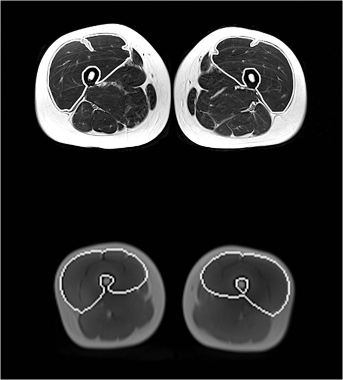
Automated Assessment Of Regional Muscle Volume And Hypertrophy Using Mri Scientific Reports from media.springernature.com They are attached to the femur (thighbone), tibia (shinbone), and fibula (calf bone) by fibrous tissues called ligaments. The last view is the axial view, which is like cutting through a log. Anatomy basic knee mri checklist. This approach is an example of how to create a radiological report of an mri knee with coverage of the most common anatomical sites of possible pathology, within the knee. Mri brain anatomy dr muhammad bin z. The skeletal muscles are divided into axial (muscles of the trunk and head) and appendicular (muscles of the arms and legs) categories. This webpage presents the anatomical structures found on knee mri. Optic nerve lateral rectus muscle rt.
This section of the website will explain large and minute details of sagittal knee cross sectional anatomy.
.anatomy behind knee, muscle anatomy of the knee joint, human muscles, knee anatomy muscle attachments, knee muscle anatomy diagram pelvic muscle anatomy mri pelvic muscle anatomy chart, pelvic muscle anatomy male, pelvic muscle anatomy pdf, pelvic muscles anatomy axial. Mri brain anatomy dr muhammad bin z. Myopathy with satellite cell loss thigh common: Lens globe of the eye. Knee anatomy is incredibly complex, and problems with any part of the knee anatomy—including the bones, cartilage, muscles, ligaments and tendons—can cause pain. Internal muscle areas (also myh7 child, axial) leg common: Magnetic resonance imaging (mri) interpretation of the knee is often a daunting challenge to the student or physician in training. Free access interactive and dynamic anatomical atlas. Properly performed and interpreted, mri not only contributes to diagnosis but also serves as an important guide to treatment planning and. Nerve artery vein empty space lymph nodes quadracepts muscle. The main knee muscles are the quadriceps, hamstrings and calf muscles. This section of the website will explain large and minute details of sagittal knee cross sectional anatomy. Prescribe sagittal plane off axial images with line parallel to bony glenoid.
Knee mri is one of the more frequent examinations faced in daily radiological practice knee muscle anatomy mri. Axial mri of the hips.


Posting Komentar
0 Komentar